


Viruses as a potential environmental trigger of type 1 diabetes mellitus (Review)
- Authors:
- Published online on: March 26, 2024 https://doi.org/10.3892/br.2024.1770
- Article Number: 81
-
Copyright: © Alves Abrantes et al. This is an open access article distributed under the terms of Creative Commons Attribution License.
Abstract
1. Introduction
Type 1 diabetes mellitus (T1DM) is a chronic metabolic disorder of autoimmune nature, characterized by the selective and progressive destruction of insulin-producing pancreatic β-cells by autoreactive T cells, leading to loss of secretory function and insufficient hormone production (1-3). This immune system dysregulation is caused by the combined action of genetic, epigenetic, and environmental factors, through a complex and varied network of interactions (4,5).
In mammals, insulin is an essential anabolic hormone with multiple effects on glucose, lipid, protein, and mineral metabolism. It is required for the entry of glucose into muscle and fat cells, in addition to stimulating the liver to store glucose in the form of glycogen and to synthesize fatty acids. Insulin is also required to stimulate amino acid uptake, inhibit the breakdown of fat in adipose tissue, and stimulate the uptake of potassium by cells (6).
Given its importance, glucose metabolism needs to be strictly regulated to ensure a sufficient energy supply to vital tissues and organs, but not supply an excess. In this context, the liver plays a critical role in glucose metabolism homeostasis, regulating multiple metabolic pathways, including glycogenesis, glycogenolysis, glycolysis, and gluconeogenesis (7). Thus, the controlled expression of genes encoding the liver enzymes involved in these pathways plays a critical role in the complex network of interactions between multiple organs. Therefore, the regulation of the expression of these genes is essential for the maintenance of long-term glucose metabolism homeostasis (8).
Control of blood glucose levels depends on a complex and tightly controlled network of hormones and neuropeptides released by the brain, pancreas, liver, intestine, adipose tissue, and muscles (9,10). However, the pancreas plays a critical role within this network since the pancreatic islets contain insulin-producing β-cells and glucagon-producing α-cells. Through these two hormones, the pancreas regulates glucose metabolism, where insulin lowers blood glucose levels, while its counterpart, glucagon, helps maintain necessary blood glucose levels; thus, acting together antagonistically, they maintain energy homeostasis (9). The loss of this balance, due to the absence or dysfunction of β-cells, generates insulinogenic and hyperglycemia, which are the classic phenotypes of T1DM (11).
Evidence suggests that genetic characteristics, notably the genetics of HLA, specifically of the molecules DQ2 and DQ8, are a necessary but insufficient condition for the development of T1DM. Thus, other additional factors, including those related to environmental stress, the individual's microbiome, and the epigenome, are also important in the pathogenesis of the disease (1,12). The increase in the incidence of T1DM in recent decades cannot be explained solely based on genetic factors, although HLA-DR3-DQ2 or HLA-DR4-DQ8 haplotypes, or both, are considered risk factors for the disease (13,14).
This suggests that the origin of the disease depends on the interaction between multiple factors, involving predisposing genes and exposure to environmental factors that act as triggers. Evidence of this is that these processes are heterogeneous even among genetically related individuals, suggesting an interaction between a genetic predisposition and other non-genetic factors, including viral infections (15). Three phenomena acting in parallel contribute to the onset of T1DM: Early childhood lifestyle and factors, the dynamics of immune system development, and the maturation of the gut microbiome (16).
The potential of viral infections to trigger pancreatic islet autoimmunity in patients with T1DM has been a longstanding hypothesis. There is an increasing body of evidence that implicates persistent infections by certain viruses, particularly human enteroviruses, as more likely environmental triggers that may contribute to different stages of disease development (17).
Studies show that infections with certain viruses play a crucial role in the pathogenesis of T1DM and can determine whether a genetically susceptible individual will develop this metabolic disease (3,18). Growing evidence points to a causal relationship between infections by certain viruses, especially human enteroviruses, and disease onset. Although the participation of viruses in the etiology of T1DM has been extensively explored, definitive proof of this causality is still lacking (18,19). However, there are reports of viral infections in several individuals with autoimmunity and damage to insulin-producing pancreatic islet β-cells, in which enteroviruses are the prime suspects (20).
Studies performed in Finland show that viral effects can encompass all stages of the β-cell damage process, including those that precede the onset of the disease (18). Among the strongest candidate viruses, enteroviruses detected in the pancreas of patients with T1DM stand out. In addition, epidemiological research, including a meta-analysis of 56 studies, has shown more infections by these enteroviruses in diabetic patients than in healthy subjects (18,21,22).
Enteroviruses cause T1DM during experimental infection of animals. Epidemiological studies in humans indicate that they are associated with an increased risk of disease and have been detected in the pancreas of diabetic patients (23,24). These results provide consistent evidence that enteroviruses trigger the autoimmune response against pancreatic islets resulting in inflammation, destruction of β-cells, and initiation of T1DM (20,25). Among the enteroviruses, Coxsackie B appears to be the primary candidate for the trigger or acceleration of islet β-cell autoimmunity in genetically susceptible individuals, resulting in T1DM (26).
The present review presents and discusses the most recent advances regarding the role of viruses as an environmental trigger, contributing to the loss of immune self-tolerance and triggering autoimmunity. A literature review was conducted using the PubMed, Embase, and Scopus databases, employing the descriptors: Type 1 diabetes, pathogenesis, virus, and associated terms with the appropriate Boolean modifiers. Studies involving humans or animals, original articles, and review articles regarding meta-analysis published in recent years were included, while abstracts, conference papers, editorials, and unrelated studies were excluded.
2. Pathogenesis of T1DM
T1DM is an organ-specific autoimmune disease that leads to the destruction of insulin-producing pancreatic β-cells, where the balance between regulatory and effector T cells determines the risk of developing the disease, activation time, and duration (27). Its origin is multifactorial, and the pathogenesis is complex, involving humoral and cellular immune response abnormalities (28). The mechanisms that trigger the disease involve the production of autoantibodies against autoantigens produced by insulin-producing pancreatic islet β-cells. Although these autoantibodies do not play a significant role in the destruction of these cells, they serve as indicators of an ongoing destructive process and as a strong predictive marker of the future development of T1DM (1,29,30). Pancreatic islet β cells produce self-antigens among them: Islet cell antigens (ICA), glutamic acid decarboxylase 65-kilodalton (GAD65), insulinoma antigen-2 (IA-2), insulin antigen (IA) and zinc transporter 8 antigen (ZnT8A), which induce the production of their respective autoantibodies (31,32).
A long-term follow-up longitudinal study showed that pancreatic islet autoimmunity precedes the onset of T1DM in prediabetic children and the presence of autoantibodies does not necessarily lead to overt disease with clinical symptoms (33). The disease progresses in three predictable and identifiable sequential steps before the onset of symptoms: i) Humoral autoimmunity with the presence of autoantibodies against antigens produced by pancreatic islet β-cells but without dysglycemia; ii) cellular autoimmunity with the presence of autoreactive cells against β-cells with dysglycemia but no symptoms; iii) and the final stage is characterized by hyperglycemia and the presence of symptoms such as polyuria, thirst, hunger, and weight loss (1,34) (Fig. 1). Of note, children with autoantibodies to IA, GAD, and IA-2 showed different progression paths, and insulin autoantibodies were most correlated with the development of T1DM (33).
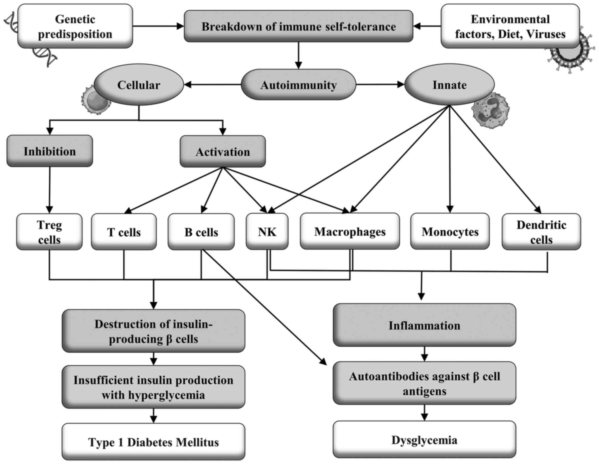
The consumption of advanced glycation end products (AGEs) may be an environmental component that together with genetic factors may play a role in the initiation of T1MD. AGEs bind to specific cell surface receptors (RAGE) and have a pro-inflammatory role (35). In the NOD mouse model, the first cells to infiltrate the pancreas are those of innate immunity, such as monocytes, natural killer cells, CD11c + dendritic cells, and ER-MP23 macrophages, with the occurrence of insulitis, self-reactive B cells and later, self-reactive T cells (27).
Humoral autoimmunity. In an observational cohort study, the majority of the patients tested positive for ≥1 type of autoantibodies against the autoantigens GAD, IA-2, and ZnT8. The presented symptoms were similar among participants with or without autoantibodies, as was the frequency of ketoacidosis. The positivity for autoantibodies decreased with age, particularly in men compared to women and non-white individuals compared to white individuals. The body mass index (BMI) was higher in adults without autoantibodies than in adults with autoantibodies. Individuals negative for autoantibodies were found to more likely to have a parent with diabetes and less likely to have another autoimmune disease (36).
The development of T1DM is preceded by an initial phase characterized by the presence of insulitis, a type of inflammatory response mediated by T lymphocytes, followed by the detection of one or more types of autoantibodies against the autoantigens produced by the β-cells of the pancreatic islets, such as IA, GAD, protein tyrosine phosphatase, IA-2 or IA-2β, and ZnT8, which are indicative of the immunological onset of T1DM (37-39). The presence of >1 type of autoantibody marks the first stage of the disease; the second stage is marked by dysglycemia or glucose intolerance, both asymptomatic. The third stage, in turn, is defined by the clinical manifestation of T1DM with symptoms of hyperglycemia, such as polyuria, polydipsia, enuresis, weight loss, blurred vision, and sometimes ketoacidosis or diabetic hyperosmolar syndrome (37,39).
A longitudinal follow-up study of a patient who developed T1DM 23 years after the idiopathic CD4 lymphocytopenia diagnosis found that the anti-GAD antibody had already been present in low titers for at least 16 years and began to increase 6 months before the onset of illness. Seroconversion to IA and IA-2 autoantigens was detected during the onset of disease symptoms. The proportion of CD8/CD4 lymphocytes increased gradually for 8 years before the disease began, in particular CD8+ T cells autoreactive against proteins related to the catalytic subunit of glucose-6-phosphatase specific to pancreatic islets at the beginning of T1DM. The patient in this study had idiopathic CD4 lymphocytopenia and a high number of islet antigen-specific CD8+ T cells that may contribute to the autoimmune destruction of insulin-producing β-cells (40).
Seroconversion to pancreatic islet autoantibodies precedes disease onset by many years, but the role of humoral autoimmunity in the initiation and progression of T1DM is still unclear. A new autoantibody directed to extracellular epitopes of zinc transporter 8 antigen (ZnT8ec), expressed in pancreatic islet cells, was identified in newly diagnosed patients with the disease, detected by immunofluorescence. When T1DM patients were compared with healthy controls the positivity rate for the antibody (ZnT8ecA) was 23.6% (41). The sequential expression of the autoantibodies: IAA, GADA, IA-2A, and ZnT8ecA was evaluated in a group of 30 children in a longitudinal follow-up study from the evolution to the clinical manifestation of T1DM, and 10 of them were positive for ZnT8ecA. Notably, ZnT8ecA was the first antibody to appear in all 10 children (41).
Glutamate is the primary excitatory neurotransmitter and is inactivated by cellular uptake via subtypes of glutamate transporters GLT-1 (EAAT2) and GLAST (EAAT1) (42). It has been shown that GLT1/EAAT2 is expressed in the membrane of pancreatic islet β-cells and regulates extracellular glutamate concentrations, preventing induced β-cell death. Patients with T1DM had autoantibodies against β-cells, but the target antigen and pathogenic mechanisms are unknown (43,44). It was hypothesized that GLT1 could be the target of autoantibodies and may be associated with the pathogenesis of T1DM. ELISA showed that sera from individuals with T1DM recognized GLT1 expressed in the brain and pancreatic islets of mice and COS7 cells transfected with the GLT1 gene. These findings were validated in two cohorts of patients with T1DM by immunofluorescence assays detecting autoantibodies against GLT1 in 37% of subjects with the disease and none of the healthy controls (45). Furthermore, in the absence of complement, autoantibodies against GLT1 markedly reduced the activity of this transporter in βTC3 cells, inducing the internalization of GLT1, leading to the death of β-cells. Thus, GLT1 is a novel T1DM autoantigen and anti-GLT1 autoantibodies cause β-cell death in the presence and absence of complement (45).
The concentrations of autoantibodies GADA and IA2A showed no statistical difference between the two groups, but the positivity rate for ZnT8A was higher in the group with ketoacidosis (46,47). ZnT8A-positive patients had higher IA2A titers and a higher prevalence of GADA and IA2A compared to ZnT8A-negative patients. Multivariate logistic regression analysis showed that positivity for ZnT8A was associated with an increased risk of microalbuminuria independent of age, sex, and BMI. This study concluded that the autoantibody, ZnT8A, had diagnostic value for ketoacidosis in children with T1DM, showing greater specificity than the other two autoantibody types. In addition, positivity for ZnT8A was related to a higher titer of IA2A and a higher frequency of multiple diabetes-related autoantibodies, a risk factor for T1DM, independent of microalbuminuria. Thus, positivity for ZnT8A may be related to ketoacidosis and microalbuminuria, accelerating the progression of T1DM (48).
A case-controlled study involving 20 patients with recent-onset T1DM, with six months or less of diagnosis and testing positive for ≥1 islet autoantibodies and 20 healthy controls negative for islet autoantibodies found that 474 genes were differentially expressed in individuals with T1DM, most of which were related to host defense, inflammatory, antibacterial, and antiviral effects, and cell cycle progression. Conversely, downregulated genes were involved in T1DM target cell repair, inflammation control, and immune tolerance. Among the genes were AREG, which is related to the expression of FOXP3, and the SMAD6 gene which is associated with immunological tolerance. SMAD6 expression was found to be negatively correlated with islet ZnT8 autoantibody. PDE12 gene expression, which resists viral pathogens, was reduced in T1DM and negatively related to the ZnT8A and GADA autoantibodies levels. Conversely, diabetic patients showed increased expression of genes encoding long non-coding RNAs, MALAT1 and NEAT1, which are related to inflammatory mediators, autoimmune diseases, and innate immune response against viral infections (49).
Cellular autoimmunity. Cellular immunity plays a critical role in the selective and progressive destruction of insulin-producing pancreatic islets β-cells, with a drastic reduction in the production of this hormone and, consequently, in the pathology of T1DM. Evidence of this is the presence of infiltration of TCD4+ and TCD8+ lymphocytes, B lymphocytes, natural killer (NK) cells, dendritic cells (DCs), macrophages, and other autoreactive immune cells that recognize pancreatic islets as foreign, resulting in their destruction, and establishment of the clinical disease (1,29,50).
CD8+ T cells are responsible for the autoimmune destruction of insulin-producing β-cells. Rapid disease progression is associated with an increased number of islet-specific CD8+ T cells with a transitional memory phenotype (51). They analyzed the phenotype and function of these cells in the progression of T1DM and identified islet-specific CD8+ T cells by high-content single-cell mass cytometry in combination with peptide-loaded Human Leukocyte Antigen (HLA) tetramer staining. A novel analytical method, DISCOV-R, was used to characterize rare subsets of CD8+ T cells. Autoreactive T cells are phenotypically heterogeneous and differ as a function of the rate of disease progression. Activated islet-specific memory CD8+ T cells were prevalent in T1DM patients with a rapid loss of C-peptide. In contrast, the slow progression of the disease is correlated with a profile of exhaustion and expression of multiple inhibitory receptors, reduced production of cytokines, and low proliferative capacity. Thus, the correlation between these properties of autoreactive CD8+ T cells and the rate of progression of T1DM after its onset can be considered an attractive phenotypic biomarker of the disease trajectory (52).
The class I HLA-B*3906 and HLA-A*2402 genes are associated with a higher risk of T1DM and promote the early onset of the disease, suggesting that CD8+ T cells that recognize the peptides presented in the context of these class I molecules on cells β play a major role in the autoimmune response that results in disease (53). A study analyzed the frequency and phenotype of CD8+ T cells specific for the β-cell antigens: Pre-proinsulin (PPI) and circulating B-specific insulin (InsB) in HLA-B*3906+ children newly diagnosed T1DM and in high-risk HLA-A*2402+ children prior to the appearance of disease-associated autoantibodies and before the diagnosis as T1DM. Antigen-specific CD8+ T cells were detected using these HLA class I tetramers, and memory status was assessed by flow cytometry. HLA-B*3906+ children with T1DM had an increase in memory CD8+ T cells specific for the preproinsulin epitope PPI5-12 compared to healthy subjects, matched by age and HLA genotype. In high-risk HLA-A*2402+ children, the percentage of terminal effector cells within InsB15-24-specific CD8+ T cells increased before diagnosis compared to samples collected before the appearance of autoantibodies. These results indicate that CD8+ T cells autoreactive to β-cells restricted by disease-associated HLA class I molecules exhibit an experienced phenotype to antigens and show enhanced effector function during the period leading up to clinical diagnosis (54).
The population of CD8+ T cells specific for β-cell antigens in T1DM maintains its self-reactive potential despite having access to a permanent source of these antigens. The longevity of these cells was evaluated, and the T-cell multipotency index was established based on DNA methylation levels. β-cell-specific CD8+ T cells maintained a stem-like epigenetic multipotency score. Single-cell assay for transposase-accessible chromatin using sequencing revealed the coexistence of epigenetic programs of naive T cells associated with those of individual β-cell specific effector CD8+ T cells. Analysis of the anatomical distribution of β-cell-specific CD8+ T cells and the setting of stem-associated epigenetic programs showed that self-reactive CD8+ T cells isolated from mouse lymph nodes maintained developmentally plastic phenotypic and epigenetic profiles relative to the same cells isolated from the pancreas. These data point to a new vision into the lifespan of β-cell-specific CD8+ T cell responses (55).
Analysis of gene expression by RNA sequencing in CD4+ and CD8+ T cells, natural killer (NK) and B cells, and chromatin accessibility by transposase accessible chromatin sequencing assay (ATAC-seq) occurred. Gene expression was evaluated in five genetically at-risk children with islet autoantibodies, with a median age of 3 years, who progressed to T1DM and compared with five sex, age, and HLA-DR-matched children who did not develop the disease. In children with the disease, the differentially expressed genes (DEGs) were mostly confined to CD4+ T cells and enriched for genes and pathways linked to cytotoxicity (56). Several highly expressed DEGs were validated in a semi-independent cohort of 13 children who progressed to the disease and 11 who did not. Flow cytometry revealed that disease progression was associated with the expansion of CD4+ cells with a cytotoxic phenotype. The ATAC-seq method showed that progression to T1DM was associated with the reconfiguration of chromatin regulatory regions in CD4+ cells, some of them linked to differentially expressed genes associated with cytotoxicity. These findings suggest that cytotoxic CD4 T cells play a role in the progression of T1DM (56,57).
Autoimmune diseases mediated by CD8 T cells result from the breakdown of self-tolerance mechanisms in self-reactive CD8 T cells (58). In T1DM, there is a loss of tolerance to pancreatic islet autoantigens due to defects in both central tolerance, which does not eliminate potentially self-reactive lymphocytes, and peripheral tolerance, which fails to control self-reactive T cells that have escaped the thymus. This breakdown of the self-tolerance mechanisms of β-cell-specific self-reactive CD8 T cells results in the destruction of these insulin-producing cells (27,59).
To understand the mechanism by which this occurs, the fate of β-cell-specific CD8 T cells was analyzed in non-obese diabetic (NOD) mice. A stem-like autoimmune progenitor population was identified in the pancreatic draining lymph node that self-renewed giving rise to autoimmune mediators that migrate to the pancreas, where they further differentiate and destroy β-cells. The transfer of only 20 cell autoimmune progenitors induced T1DM, while up to 100,000 autoimmune pancreatic mediators could not. Pancreatic autoimmune mediators are short-lived, stem-like autoimmune progenitors that must continually seed the pancreas to sustain β-cell destruction. Single-cell RNA sequencing and clonal analysis revealed that autoimmune CD8 T cells are unique T cell differentiation states with identified characteristics that suggest the transition from autoimmune progenitor to autoimmune mediator (60).
The molecule CD137 is a member of the tumor necrosis factor receptor family and modulator of T1DM progression in NOD mice. CD137 expression on CD4 T cells inhibits the development of the disease, but on CD8 T cells it promotes T1DM, increasing CD8 T cells self-reactivity to β-cells (61). CD137 is expressed in a subpopulation of FOXP3+ CD4 regulatory T cells (Tregs), which constitutes the primary source of soluble CD137 that suppresses T cells, binding to its ligand, CD137L, whose expression is increased on activated T cells. NOD mice transfected with the CD137L gene (NOD.Tnfsf9-/-) significantly delayed the development of T1DM, had less insulitis, and had a reduced quantity of CD8 T cells autoreactive to β-cells when compared to wild-type NOD mice. Adoptive T cell transfer showed that CD137L deficiency on myeloid APCs was associated with T1DM suppression. Conversely, the lack of CD137L in T cells increased their diabetogenic activity. Neither CD137 nor CD137L is required for the development and homeostasis of FOXP3+ Treg cells. However, CD137 was critical for T1DM suppressive activity in vivo by FOXP3+ Treg cells, suggesting that the interaction between CD137 and CD137L regulates this function (62).
Regulatory CD4+ T cells (Tregs) act as protectors against T1DM, recognizing antigens such as insulin or pancreatic islet peptides, presented in the context of HLA-DQ6 or DR15, which are protective haplotypes against diabetes (63). This interaction generates the production of anti-inflammatory cytokines, such as IL-10 and TGF-β and this activation of antigen-specific Tregs protects β-cells from destruction through four mechanisms: i) Damage to cells occurs when CD4+ T cell effectors recognize antigens of islet cells presented by APCs, in the HLA-DQ8 or DR4 context, which is a risk for T1DM, and are activated and migrate to the pancreas, where they induce β-cell destruction (64). Antigen-specific Tregs act by suppressing effector CD4+ T cells and preventing their migration to the pancreas through the release of anti-inflammatory cytokines. ii) Effector CD8+ T cells in pancreatic islets recognize self-antigens presented in the HLA-A2 context by β-cells and destroy these cells. Tregs can suppress effector CD8+ T cells and prevent β-cell destruction by releasing anti-inflammatory cytokines. iii) Tregs can reduce antigen-independent granzyme-mediated APC death. iv) Alternatively, Tregs can induce antigen-dependent APC death when the antigen is presented to the T cell receptor (TCR) of Tregs (63).
3. Role of viruses in the development of T1DM
The rapid growth in the incidence of T1DM suggests that environmental factors, including viruses, play a significant role in the pathogenesis of the disease. Some microbial agents may act as risk or protective factors for the disease. To date, two hypotheses have attempted to explain these effects: The hygiene hypothesis suggests that exposure to these agents in early childhood stimulates immunoregulatory mechanisms that protect against autoimmunity. The triggering hypothesis suggests that specific agents can damage insulin-producing β-cells and contribute to autoimmunity (23,65,66). Certain viruses, particularly enteroviruses, are the primary suspects and candidates for risk factors of T1DM. According to epidemiological studies, they can cause disease in animals and are associated with an increased risk of diabetes in humans, in addition to being detected in the pancreas of patients with T1DM. The possible protective effect of certain microbes has been investigated in animal models and epidemiological studies, in which certain enteric agents and gut microbiome patterns were associated with a reduced risk of T1DM (20,23).
Growing evidence continues to implicate enteroviruses as the most likely triggering agents of autoimmunity in T1DM patients, possibly through persistent infections, contributing to different stages of the disease (17,67). A meta-analysis included 38 studies covering 5,921 subjects from all continents, including 2,841 T1DM patients and 3,080 healthy controls. Pooled analysis showed that enterovirus infection was positively associated with disease in European, African, Asian, Australian, and Latin American populations, but inconclusive for North America. This association between enterovirus infection and T1DM was found in blood and tissue samples, but no association was observed in stool samples (68). Among the viruses that may be associated with an increased risk for T1DM, enteroviruses, primarily Coxsackie, echoviruses, in addition to rotavirus and, to a lesser extent, mumps, parainfluenza, rubella, and cytomegaloviruses stand out (20,23,68,69).
The long-term interaction between genetic, epigenetic, and environmental factors, including viral infection, can increase or decrease an individual's likelihood of developing an autoimmune disease, depending on the imbalance between risk factors and protective effects; three main mechanisms have been proposed to explain the development of autoimmunity in T1DM: molecular mimicry, epitope spread, and bystander activation (70).
Studies report the association between viral infections and the development of T1DM, but the immunological mechanisms involved and the link between viral infections and disease onset or progression are still unclear (71). One of the most commonly discussed possibilities is molecular mimicry which involves cross-reactive immunity against epitopes shared between viruses and human pancreatic β-cells. Thus, molecular mimicry may cause the activation of self-reactive T cells in T1DM, activated by a virus that carries an epitope bearing a strong similarity to certain structures present in human β-cells (72). This triggers a cross-reactive autoimmune response that eliminates the viral infection and the β-cells of the pancreatic islets. This mechanism may explain how certain viruses may play a role in triggering T1DM (71,73).
Certain viruses infect and damage pancreatic β cells, whose innate antiviral immune response can be modulated by specific viral RNA receptors and sensors, including the melanoma differentiation-associated gene 5 (MDA5), encoded by the IFIH1 gene (74). MDA5 has been specifically linked to inflammation and cell death of pancreatic cells from rotavirus-infected mice models (75). Activation of the MDA5 receptor limits rotavirus infection through the induction of the interferons and pro-inflammatory cytokine production but may also facilitate progression to an autoimmune response with the destruction of pancreatic β-cells. Polymorphisms in the IFIH1 gene, which encodes MDA5, are associated with an increased risk of T1DM (76).
MDA5 receptor activation is associated with inflammatory and immunoregulatory responses from pancreatic islets to viral infections and is expressed in pancreatic endocrine cells with preferential localization to α-cells compared to β-cells, suggesting that α-cells are better equipped than β-cells to respond to viral infections and initiate viral clearance mechanisms. This difference appears to make β-cells more susceptible to the establishment of persistent low-grade viral infections. MDA5 is increased in patients with new-onset T1DM, possibly because of elevated inflammatory conditions (77).
Growing evidence shows that the post-translational processing of peptides may play a role in the immune response under pathological conditions by molecular mimicry. This mechanism appears particularly relevant in T1DM, as epitopes resulting from post-translational splicing derived from disease-related antigens linked to HLA class I and II complexes. CD4+ and CD8+ T cell-mediated immune responses in T1DM patients confirmed its immunogenicity (78). Peptide processing theoretically generates large sequencing variability, increasing the frequency of human-viral zwitter peptides, which share complete sequence homology, regardless of whether they originate from human or viral antigens, impairing discrimination between self and non-self-antigens by T cells, increasing the risk of virus-triggered autoimmune responses (79).
Linear sequence similarities in amino acid motifs are not the only criterion for developing molecular mimicry autoimmunity. Autoreactive immune cells are prepared by molecular mimicry, becoming sensitized but not causing apparent disease. However, subsequent environmental insults can induce these previously sensitized autoreactive cells to induce autoimmune disease (80). The association between viral infections and autoimmune diseases has long been reported as events that precede target organ inflammation (81). Virally-infected mice that were later challenged following viral shedding, with a nonspecific immune insult developed autoimmune disease (82). Conventional inflammatory responses to specific pathogens induced autoimmune disease in animals primed with a molecular mimic for a CNS antigen (83). Therefore, activation of the immune system alone is not a necessary condition for autoimmune disease, but the environment in which sensitized immune cells are exposed to antigens is an important factor in the onset of the disease (80).
During viral infections, a significant quantity of T cells are activated in a T cell receptor (TCR)-independent and cytokine-dependent manner, by a mechanism known as ‘bystander activation’. Type I interferons, IL-18, and IL-15, are the most important factors that induce bystander activation of T cells, each of which plays a somewhat different role (84). Bystander-activated T cells do not have specificity for the pathogen but can affect the course of the immune response to an infection. For example, Bystander-activated CD8+ T cells participate in protective immunity by secreting cytokines, such as interferon-γ. Conversely, they also cause damage to the host by exerting cytotoxic effects facilitated by NK cell activation receptors, such as NKG2D, and cytolytic molecules, such as granzyme B. Interestingly, there is a strong association between the cytolytic function of bystander-activated CD8+ T cells and liver injury in patients with acute infection with the hepatitis A virus (84,85).
Bystander activation occurs when CD8+ and CD4+ T cells are activated in an antigen-independent manner in the absence of TCRs. This alternative mechanism encompasses activation through soluble factors or membrane-bound molecules that bind to receptors other than the TCR (84). In certain viral infections, this activation is beneficial for clearing the virus but also triggers the activation of autoreactive T cells in individuals genetically predisposed to autoimmunity (86). Memory T cells are the most vulnerable to bystander activation as they express cytokines and co-signaling receptors. This activation occurs due to an inflammatory environment, co-signaling ligands, and interactions with neighboring cells (70).
Several types of T cell populations, such as NKT, and Tγδ, in addition to conventional CD4+ and CD8+ T cells, can be induced to exhibit innate-type effector function through bystander activation (86,87). Antigen-specific T cells are the hallmark of the adaptive immune response, while non-antigen-related T cells in an inflammatory environment proliferate and synthesize effector cytokines. In a viral infection, antigen-specific T cells are activated by cytokines to ensure protective immunity. Bystander activation of various types of T cells in the absence of antigen recognition with the production of inflammatory cytokines may also contribute to the elimination of a pathogen. Conversely, the function of bystander-activated T cells may also be a mechanism in the pathogenesis of autoimmune disease. Studies have reported that non-antigen-related T cell infiltration into inflammatory tissues participates in an autoimmune response (87).
Role of enteroviruses. Human enteroviruses are small, non-enveloped, positive-sense, single-stranded RNA viruses belonging to the Picornaviridae family and the Enterovirus genus, which includes 7 species involved in human diseases, including Poliovirus and Coxsackie, which constitute five groups: The A group with 24 serotypes, the B group with 6 serotypes, the C group with 23 serotypes, the D group with 5 serotypes and the Echoviruses group (88,89). They are responsible for several human infections, ranging from asymptomatic to mild and severe diseases, as well as certain chronic autoimmune diseases, including T1DM (25).
Although they are highly cytolytic, enteroviruses can persist in various tissues, including pancreatic islets, and this persistence is hypothesized to play a role in the pathogenesis of T1DM. Viral persistence is the result of virus-host coevolution leading to cellular resistance to lysis due to mutations or downregulation of the viral receptor and reduced replication (90). Group B coxsackieviruses are more commonly implicated in the development of T1DM. Its persistence in pancreatic cells can trigger autoimmunity in genetically predisposed individuals, destroying insulin-producing β-cells through the activation of inflammation. The persistence of this virus in the intestine, blood cells, and thymus has been reported and possibly contributes to a certain extent to the pathogenesis of T1DM (26,90).
Environmental factors can initiate and possibly sustain, accelerate, or delay pancreatic β-cell damage. The role of these factors has been extensively studied, and viruses, especially enteroviruses, stand out as the most likely candidates. The detection of enteroviruses in T1DM patients in randomized trials confirmed the importance of human enteroviruses in the pathogenesis of the disease (17). Genetic susceptibility and innate and acquired immune responses against enteroviruses may somehow contribute to the breakdown of tolerance to self-antigens. The frequency with which this occurs, the mechanisms and pathways of activation of virus-induced autoimmunity, and the destruction of β-cells in T1DM have yet to be determined (91).
The detection of enteroviruses in the serum, blood, feces, nasal swabs, and pancreatic tissue of patients with T1DM, before or close to the clinical onset of the disease, suggests an association between infections with these viruses and the disease (24). However, definitive evidence of the role of these pathogens in the onset and progression of the disease is still lacking. Emerging evidence suggests that chronic human enterovirus infections occur in the pancreas of patients with T1DM. However, the lack of sensitive molecular techniques capable of detecting low quantities of viral proteins and RNA remains an obstacle. Enterovirus RNA has been frequently detected in various tissues of patients with T1DM and the presence of the virus in pancreatic islets suggests persistent slow-replicating infection that may have a role in the autoimmune response, which results in T1DM (24,92).
Functionally, the thymus can be compared to a computer highly specialized in orchestrating central immune self-tolerance, a necessary condition for the survival of the species, from the evolutionary pressure imposed by the hostile environment and the autotoxicity inherent to the stochastic generation of the diversity of immune cell receptors that characterize the adaptive immune response. The presentation of self-antigens in the thymus is responsible for the clonal deletion of autoreactive T cells, which arise during the random recombination of gene segments that encode variable parts of the TCR. At the same time, the presentation of autoantigens in the thymus generates Tregs that can inhibit, in the periphery, the autoreactive T cells that have escaped negative selection in the thymus. Thus, the autoimmunity against neuroendocrine glands is due to a defect in intrathymic programming breaking the immune self-tolerance that can be genetic or acquired during, for example, an enteroviral infection (93).
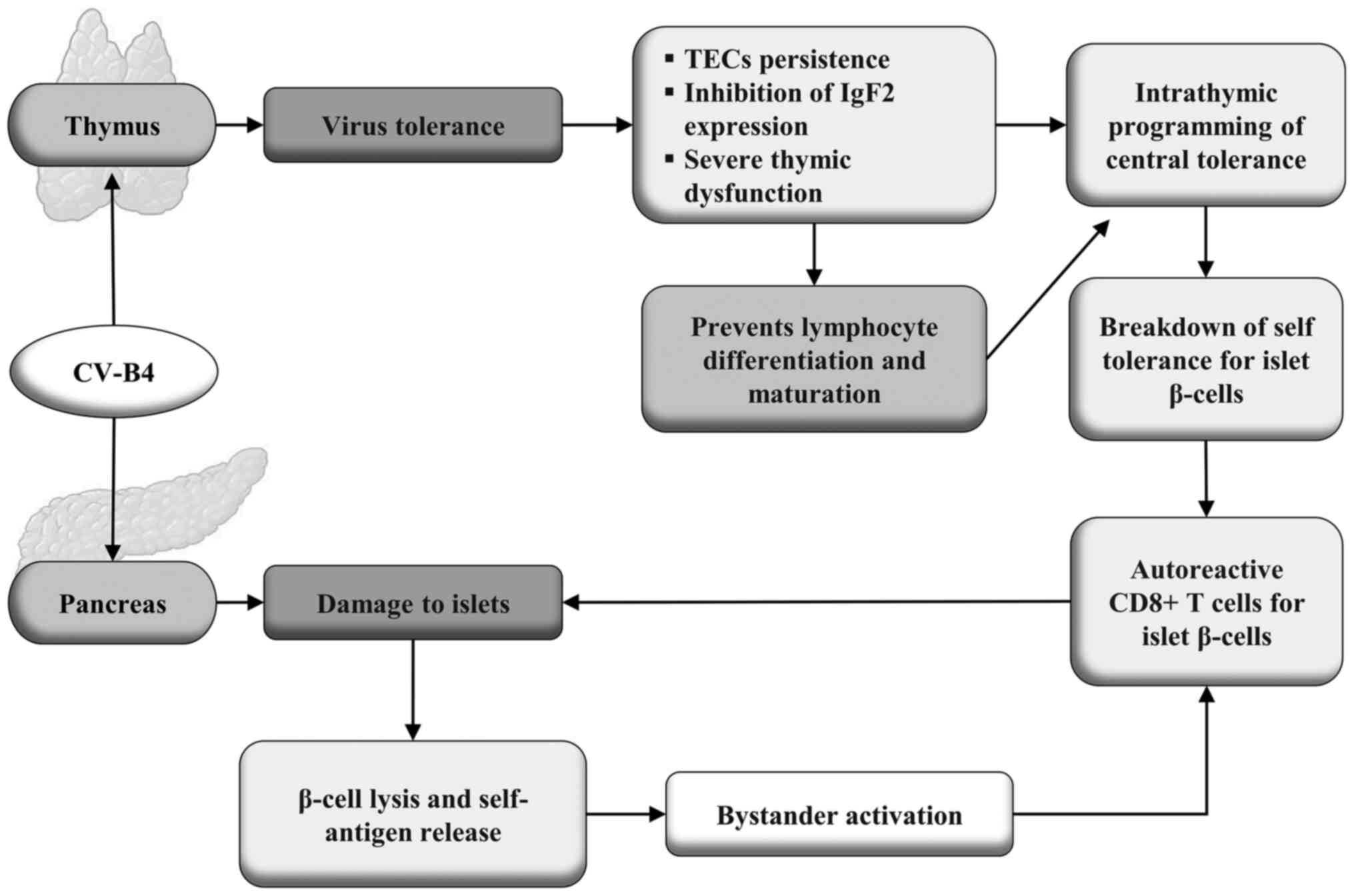
Whether or not viruses can disrupt thymus function and play a role in the pathogenesis of autoimmune diseases is an open question (94). It is known that through the synthesis and presentation of HLA-linked neuroendocrine autoantigens, thymic epithelial cells (TECs) play a crucial role in programming central immune self-tolerance to neuroendocrine functions (93). Insulin-like growth factor-2 (IGF-2) is the dominant polypeptide of the insulin family expressed in these cells in different animal species and humans. Thymic infection by coxsackievirus B4 (CV-B4) may compromise the thymus function and interfere with the intrathymic programming of tolerance to insulin antigen family and secondarily to insulin-secreting pancreatic islet β-cells (95). Productive thymus infection by this virus occurs after oral inoculation in mice. Infection of a murine medullary TEC line induces a significant decrease in Igf2 gene expression and IGF-2 production. It is hypothesized that inhibition of Igf2 expression in infected TECs can lead to a breakdown of central immune self-tolerance. This leads to the triggering of autoimmune responses against self-antigens produced by insulin-secreting pancreatic islet β-cells (96).
Thymus gland dysfunction can be a crucial step in organ-specific autoimmune diseases such as T1DM. Viruses can disrupt thymic functions by inducing thymic atrophy, dysfunction, apoptosis, and impaired lymphocyte maturation (97). Enteroviruses have been widely associated with the pathogenesis of T1DM due to enterovirus-induced thymic dysfunction in natal or perinatal life. Infection of the thymus by enteroviruses can interfere with T cell maturation or lead to the production of autoreactive T cells, both being potentially involved in the development of T1DM. It has been shown that CV-B4 infection can have multiple effects on the thymus resulting in the dysregulation of tolerance involved in the pathogenesis of T1DM, but the specifics require further study (95). The possible mechanisms by which CV-B4 can trigger the autoimmune response that leads to T1DM are shown in Fig. 2.
It has been suggested that only a minority of pancreatic β-cells appear to be infected at any one time. Furthermore, enteroviruses can become defective and stop replicating, which may explain why they are rarely isolated from pancreatic tissues. It appears that enteroviral infection of β-cells largely depends on the innate and adaptive immunity of the host. Thus, viruses may play a role in T1DM at multiple levels, including stimulating chronic autoimmunity and generating inflammation that leads to β-cell dysfunction or destruction. This stressful situation contributes to autoimmunity, creating a vicious circle (98).
The Environmental Determinants of Diabetes in the Young (TEDDY) study is the largest multicenter prospective study of young children with genetic susceptibility to T1DM that aimed to identify the environmental causes of the disease. Metagenomic sequencing of monthly stool samples paired with healthy controls collected from newborns until the detection of islet autoimmunity or T1DM was performed (99). Enterovirus B was the only virus identified in the human virome with a significant association with islet autoimmunity. Although there was no difference between the frequencies between cases and controls, children with prolonged excretion of enterovirus B were more likely to develop T1DM. Interestingly, human mastadenovirus C infection early in life was less frequent in children who developed islet autoimmunity than in those who did not, suggesting a protective effect. An association between a polymorphism in the coxsackie virus and the adenovirus receptor human gene with susceptibility to T1DM led the authors to propose that competition for receptor binding between the adenovirus and the coxsackie virus conferred the protective effect of the adenovirus (69).
The coincidence of periodic epidemiological patterns of incidence of infections by enteroviruses and T1DM stimulated the first suspicions of the association of these viruses with the onset of the disease (100). Furthermore, there is a strong correlation between genetic susceptibility to islet autoimmunity and enterovirus infection. Clinical and epidemiological findings point to the coxsackie B virus as a potential triggering mechanism of an autoimmune reaction against β-cells, leading to an increase in inflammation, destruction of these cells, and T1DM development (25). Conversely, NOD mice immunized with a vaccine against coxsackievirus Bs were protected from the accelerated onset of the disease without causing inflammation of the pancreatic islets and preventing the progression of the disease induced by this virus in the animal model that develops the disease spontaneously (101).
Several serotypes of group B coxsackieviruses result in chronic infections in human cells both in vivo and in vitro, but the mechanisms leading to the persistence of these enteroviruses and autoimmunity against pancreatic β-cells are still not fully understood (102). A carrier-state-type persistent infection model using a PANC-1 human pancreatic cell line and two different coxsackie B strains was used to assess virus-induced changes in the cell transcriptome. Clear changes were observed in the gene expression of factors associated with the pancreatic microenvironment, such as the secretory pathway and lysosomal biogenesis during persistent infection. In addition, the antiviral response pathways were activated differently by the two viral strains. It was revealed that there were extensive transcriptional responses in persistently infected pancreatic cells resulting in notable alterations, with some opposing and some similar changes between the two strains (103).
A study in Taiwan showed that the incidence of T1DM was significantly higher in children aged 0-6 years old, decreasing in adolescents aged 13-19 years old. The risk of developing the disease in children aged 0-6 years infected with an enterovirus was significantly higher than in uninfected individuals. Furthermore, the incidence of T1DM in children aged 0-6 years old was positively correlated with isolation rates of the Coxsackie virus A species, but no association after 7 years of age (104). The complex interactions of epigenetic and environmental genetic factors, especially the viruses, can trigger autoimmune reactions against autoantigens produced by β cells responsible for destroying these cells, and thus, the subsequent development of T1DM. Growing evidence continues to implicate persistent enteroviral infections as the most likely potential environmental trigger for T1DM. Consistent evidence also suggests that enteroviral infections may contribute by acting at different stages of T1DM development (17).
The NOD female mouse model is suitable for studying T1DM since these mice spontaneously develop the disease, and this can be accelerated by certain viruses. Toll-like 3 (Tlr3-/-) and wild-type (Tlr3+/+) knockout female NOD mice were used to assess the role of this receptor in triggering T1DM. Islet samples from Tlr3+/+ and Tlr3-/-animals infected and uninfected with CV-B4 were analyzed by immunostaining, laser capture microdissection, and real-time reverse transcription-quantitative PCR. Tlr3+/+ mice showed a higher incidence of insulitis and expression of CXCL10, IL1β, TNFα, and TGFβ1 compared with Tlr3-/-. After CV-B4 infection, NOD Tlr3+/+ mice had a higher incidence of insulitis and T cell infiltration 3 days post-infection compared with Tlr3-/-mice also infected with CV-B4. The results indicated that the Toll-like receptor 3 was required to create an inflammatory microenvironment of pancreatic islets, increasing insulitis and the expression of cytokines that favor the development of CV-B4-induced T1DM in female NOD mice (105).
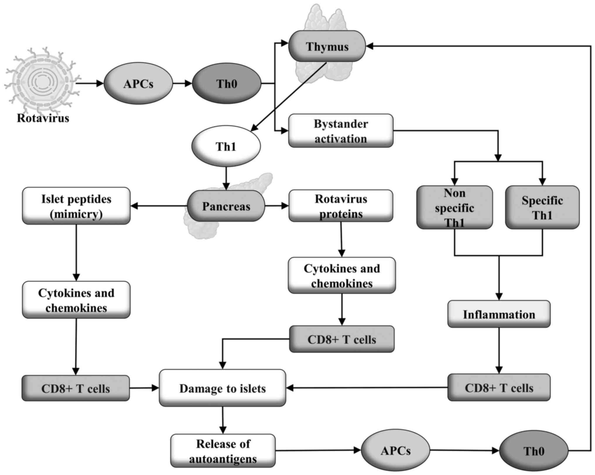
Role of rotaviruses. Rotaviruses have long been implicated as a potential environmental agent that triggers T1DM. The proposed mechanism involves molecular mimicry between the external capsid antigen of human rotavirus, VP7 protein, and the autoantigen tyrosine phosphatase IA-2 of the pancreatic islets, the molecular target of autoimmunity in T1DM. The major epitope of the intracytoplasmic domain of the IA-2 antigen has been shown to share 56% homology with a sequence of the rotavirus VP7 protein (106). The role of rotavirus VP7 protein in accelerating diabetes has also been demonstrated experimentally in NOD mice infected with rhesus monkey rotavirus (107,108).
Rotavirus and other viruses have historically been considered one of the environmental factors that trigger T1DM (108). A positive association was found between seroconversion to rotavirus VP7 protein and the increase in autoantibodies to GAD, IA-2, and insulin antigens associated with T1DM; thus, rotavirus infection is a trigger for the development of pancreatic islet autoimmunity in genetically predisposed children. A later study reinforced the theory of molecular mimicry between the rotavirus protein VP7 and the pancreatic islet peptides, IA2 and GAD5, demonstrating how the same epitope sequences in these peptides bind to T1DM-associated HLA-DRB1*04 molecules and can induce T cell proliferative responses (109,110).
Rotavirus infection generates activated immune cells with a Th1 phenotype that migrates to the pancreas where it can trigger autoimmunity through two mechanisms: i) molecular mimicry in which cross-reactive Th1 cells that recognize both the rotavirus and host peptide results in the release of cytokines and chemokines, which recruit and activate cytotoxic T cells that damage the islets resulting in the subsequent release of islet antigens that activate APCs, which then feedback in to the autoimmune process. ii) Bystander activation of nonspecific autoreactive Th1 cells and rotavirus-specific Th1 cells leads to inflammation, increasing infiltration of cytotoxic T cells and destruction of islets promoting the release of autoantigens, which activates autoreactive Th1 cells by a TCR-dependent mechanism. The self-reactive T cells activated in this manner mediate islet damage by releasing more antigens that feedback on the autoimmune response (108,111). In Fig. 3, a proposal showing the possible mechanisms by which rotaviruses trigger the autoimmune response that leads to T1DM is shown.
Rotavirus infections were initially identified as possible triggers for the initiation of T1DM due to the similarities between viral peptide sequences and human pancreatic islet peptide sequences in carriers of the disease (111). Furthermore, rotavirus infection increases the risk of T1DM in NOD mice. Research on the association between rotavirus infections with the risk of the disease in humans has produced mixed results and suggested the involvement of other factors, such as age and diet. With the global availability of rotavirus vaccines, studies have evaluated whether rotavirus vaccination alters the incidence of T1DM and have found no associations, protective or otherwise. These studies suggest a possible etiologic relationship between certain wild-type rotavirus infections and T1DM (110).
Epidemiological and immunological data suggest a strong link between rotavirus infection and two high-incidence autoimmune pathologies: Celiac disease (CD) and T1DM. The role of current oral rotavirus vaccines is being elucidated, with a positive protective association against these autoimmune diseases so far demonstrated (108), highlighting the potential role of rotaviruses as triggers of T1DM. Two recent studies suggest that the complete routine vaccination schedule against rotavirus decreased the incidence of T1DM in children. The first was performed in Australia (112). The second was a longitudinal cohort study involving 1,474,535 children in the United States, conducted by Rogers et al (113), from 2001 to 2017, using data from a health insurer in the country. Participants were classified as vaccinated when they received the series of one of the rotavirus vaccines (three doses of RotaTeq or two doses of Rotarix), or at least one dose without completing the series; as unvaccinated if they had not received any of the vaccines, and those born before its existence. Children who completed the rotavirus vaccine series had a 33% reduced risk of T1DM compared to unvaccinated children. Those who completed the RotaTeq pentavalent vaccine series had a 37% lower risk of developing the disease. Partial vaccination was not associated with the incidence of T1DM. The authors concluded that rotavirus vaccination is associated with a reduction in T1DM incidence, and that rotavirus immunization may be the first practical measure for prevention of T1DM (113). However, a less comprehensive cohort study involving 880,629 children found no evidence that rotavirus vaccination prevented T1DM (114).
In addition to viruses from the Picornaviridae family, which includes enteroviruses, such as Coxsackievirus B, and from the Reoviridae family, including Rotavirus, other viruses belonging to different families, such as the Togaviridae family (for example Rubella virus), the Paramyxoviridae family (for example Mumps virus), the Herpesviridae family (for example Cytomegalovirus and Epstein Barr virus), and the Coronaviridae family (for example SARS-CoV-2), have been identified as potential environmental factors associated with the risk of developing T1DM (3).
4. Conclusions
Viral infections are potential candidates as environmental triggers of T1DM. However, certain aspects require further clarification, such as the absence of generalized cytopathic effects on the pancreas of patients with the disease. Additionally, there is no closely related temporal association between such infections and disease onset, although there is strong evidence that at least some viruses act as environmental triggers in T1DM. The exact mechanisms by which this occurs remains to be definitively proven. Furthermore, viruses have rarely been isolated from the pancreas of individuals with T1DM, possibly due to the inaccessibility of the organ. Data available in the literature point to the role of viruses, notably enteroviruses, in the etiopathogenesis of the disease, especially during possible persistent or chronic recurrent infections of pancreatic β-cells. The detection, with a certain frequency, of RNA and viral proteins in the pancreatic tissues of individuals with T1DM represents strong evidence of latent infection or slow replication of these viruses. However, the mechanism by which they act to trigger the autoimmune process that leads to selective and progressive destruction of insulin-producing pancreatic β-cells, with disease initiation and progression, is still unclear.
Acknowledgements
Not applicable
Funding
Funding: This study was financed in part by the Coordenação de Aperfeiçoamento de Pessoal de Nível Superior-Brasil (CAPES) (Finance Code, 001).
Availability of data and materials
Not applicable.
Authors' contributions
JJPA, JVF, and JVA conceived the study, performed the literature review, and drafted the manuscript. MTFO and VDA drafted the manuscript. FLF, FLB, and TAAMF critically revised the manuscript for important intellectual content. DCFL, JMGA, and VSA were involved in the formal analysis of the article included in the review. Data authentication is not applicable. All authors read and approved the final manuscript.
Ethics approval and consent to participate
Not applicable.
Patient consent for publication
Not applicable.
Competing interests
The authors declare that they have no competing interests.
References
|
Katsarou A, Gudbjörnsdottir S, Rawshani A, Dabelea D, Bonifacio E, Anderson BJ, Jacobsen LM, Schatz DA and Lernmark Å: Type 1 diabetes mellitus. Nat Rev Dis Primers. 3(17016)2017.PubMed/NCBI View Article : Google Scholar | |
|
Roep BO, Thomaidou S, van Tienhoven R and Zaldumbide A: Type 1 diabetes mellitus as a disease of the β-cell (do not blame the immune system?). Nat Rev Endocrinol. 17:150–161. 2021.PubMed/NCBI View Article : Google Scholar | |
|
Zorena K, Michalska M, Kurpas M, Jaskulak M, Murawska A and Rostami S: Environmental factors and the risk of developing type 1 diabetes-old disease and new data. Biology (Basel). 11(608)2022.PubMed/NCBI View Article : Google Scholar | |
|
de Azevedo JCV, de Medeiros Fernandes TAA, Cavalcante GA, de Medeiros IACM, Lanza DCF, de Araújo JMG, Bezerra FL and Fernandes JV: Biology and natural history of type 1 diabetes mellitus. Curr Pediatr Rev. 19:253–275. 2023.PubMed/NCBI View Article : Google Scholar | |
|
Cerna M: Epigenetic regulation in etiology of type 1 diabetes mellitus. Int J Mol Sci. 21(36)2019.PubMed/NCBI View Article : Google Scholar | |
|
Lucier J, Weinstock RS and Doerr C: Diabetes mellitus type 1 (Nursing). StatPearls Publishing, Treasure Island, FL, 2023. | |
|
Alamri ZZ: The role of liver in metabolism: An updated review with physiological emphasis. Int J Basic Clin Pharmacol. 7:2271–2276. 2018. | |
|
Han HS, Kang G, Kim JS, Choi BH and Koo SH: Regulation of glucose metabolism from a liver-centric perspective. Exp Mol Med. 48(e218)2016.PubMed/NCBI View Article : Google Scholar | |
|
Röder PV, Wu B, Liu Y and Han W: Pancreatic regulation of glucose homeostasis. Exp Mol Med. 48(e219)2016.PubMed/NCBI View Article : Google Scholar | |
|
Chadt A and Al-Hasani H: Glucose transporters in adipose tissue, liver, and skeletal muscle in metabolic health and disease. Pflugers Arch. 472:1273–1298. 2020.PubMed/NCBI View Article : Google Scholar | |
|
Zheng P, Li Z and Zhou Z: Gut microbiome in type 1 diabetes: A comprehensive review. Diabetes Metab Res Rev. 34(e3043)2018.PubMed/NCBI View Article : Google Scholar | |
|
Cohn A, Sofia AM and Kupfer SS: Type 1 diabetes and celiac disease: Clinical overlap and new insights into disease pathogenesis. Curr Diab Rep. 14(517)2014.PubMed/NCBI View Article : Google Scholar | |
|
Pociot F and Lernmark Å: Genetic risk factors for type 1 diabetes. Lancet. 387:2331–2339. 2016.PubMed/NCBI View Article : Google Scholar | |
|
Abela AG and Fava S: Why is the incidence of type 1 diabetes increasing? Curr Diabetes Rev. 17(e030521193110)2021.PubMed/NCBI View Article : Google Scholar | |
|
Zajec A, Trebušak Podkrajšek K, Tesovnik T, Šket R, Čugalj Kern B, Jenko Bizjan B, Šmigoc Schweiger D, Battelino T and Kovač J: Pathogenesis of type 1 diabetes: Established facts and new insights. Genes (Basel). 13(706)2022.PubMed/NCBI View Article : Google Scholar | |
|
Siljander H, Honkanen J and Knip M: Microbiome and type 1 diabetes. EBioMedicine. 46:512–521. 2019.PubMed/NCBI View Article : Google Scholar | |
|
Lloyd RE, Tamhankar M and Lernmark Å: Enteroviruses and type 1 diabetes: Multiple mechanisms and factors? Annu Rev Med. 73:483–499. 2022.PubMed/NCBI View Article : Google Scholar | |
|
Hyöty H: Viruses in type 1 diabetes. Pediatr Diabetes. 17 (Suppl 22):S56–S64. 2016.PubMed/NCBI View Article : Google Scholar | |
|
Alhazmi A, Sane F, Lazrek M, Nekoua MP, Badia-Boungou F, Engelmann I, Alidjinou EK and Hober D: Enteroviruses and type 1 diabetes mellitus: An overlooked relationship in some regions. Microorganisms. 8(1458)2020.PubMed/NCBI View Article : Google Scholar | |
|
Isaacs SR, Foskett DB, Maxwell AJ, Ward EJ, Faulkner CL, Luo JYX, Rawlinson WD, Craig ME and Kim KW: Viruses and type 1 diabetes: From enteroviruses to the Virome. Microorganisms. 9(1519)2021.PubMed/NCBI View Article : Google Scholar | |
|
Geravandi S, Richardson S, Pugliese A and Maedler K: Localization of enteroviral RNA within the pancreas in donors with T1D and T1D-associated autoantibodies. Cell Rep Med. 2(100371)2021.PubMed/NCBI View Article : Google Scholar | |
|
Isaacs SR, Roy A, Dance B, Ward EJ, Foskett DB, Maxwell AJ, Rawlinson WD, Kim KW and Craig ME: Enteroviruses and risk of islet autoimmunity or type 1 diabetes: Systematic review and meta-analysis of controlled observational studies detecting viral nucleic acids and proteins. Lancet Diabetes Endocrinol. 11:578–592. 2023.PubMed/NCBI View Article : Google Scholar | |
|
Kondrashova A and Hyöty H: Role of viruses and other microbes in the pathogenesis of type 1 diabetes. Int Rev Immunol. 33:284–295. 2014.PubMed/NCBI View Article : Google Scholar | |
|
Oikarinen S, Krogvold L, Edwin B, Buanes T, Korsgren O, Laiho JE, Oikarinen M, Ludvigsson J, Skog O, Anagandula M, et al: Characterisation of enterovirus RNA detected in the pancreas and other specimens of live patients with newly diagnosed type 1 diabetes in the DiViD study. Diabetologia. 64:2491–2501. 2021.PubMed/NCBI View Article : Google Scholar | |
|
Geravandi S, Liu H and Maedler K: Enteroviruses and T1D: Is it the virus, the genes or both which cause T1D. Microorganisms. 8(1017)2020.PubMed/NCBI View Article : Google Scholar | |
|
Nekoua MP, Alidjinou EK and Hober D: Persistent coxsackievirus B infection and pathogenesis of type 1 diabetes mellitus. Nat Rev Endocrinol. 18:503–516. 2022.PubMed/NCBI View Article : Google Scholar | |
|
Bluestone JA, Herold K and Eisenbarth G: Genetics, pathogenesis and clinical interventions in type 1 diabetes. Nature. 464:1293–1300. 2010.PubMed/NCBI View Article : Google Scholar | |
|
Kahaly GJ and Hansen MP: Type 1 diabetes associated autoimmunity. Autoimmun Rev. 15:644–648. 2016.PubMed/NCBI View Article : Google Scholar | |
|
Li M, Song LJ and Qin XY: Advances in the cellular immunological pathogenesis of type 1 diabetes. J Cell Mol Med. 18:749–758. 2014.PubMed/NCBI View Article : Google Scholar | |
|
Knip M, Siljander H, Ilonen J, Simell O and Veijola R: Role of humoral beta-cell autoimmunity in type 1 diabetes. Pediatr Diabetes. 17 (Suppl 22):S17–S24. 2016.PubMed/NCBI View Article : Google Scholar | |
|
Winter WE, Harris N and Schatz D: Type 1 diabetes islet autoantibody markers. Diabetes Technol Ther. 4:817–839. 2002.PubMed/NCBI View Article : Google Scholar | |
|
Winter WE and Schatz DA: Autoimmune markers in diabetes. Clin Chem. 57:168–175. 2011.PubMed/NCBI View Article : Google Scholar | |
|
Kwon BC, Anand V, Achenbach P, Dunne JL, Hagopian W, Hu J, Koski E, Lernmark Å, Lundgren M, Ng K, et al: Progression of type 1 diabetes from latency to symptomatic disease is predicted by distinct autoimmune trajectories. Nat Commun. 13(1514)2022.PubMed/NCBI View Article : Google Scholar | |
|
Insel RA, Dunne JL, Atkinson MA, Chiang JL, Dabelea D, Gottlieb PA, Greenbaum CJ, Herold KC, Krischer JP, Lernmark Å, et al: Staging presymptomatic type 1 diabetes: A scientific statement of JDRF, the Endocrine Society, and the American Diabetes Association. Diabetes Care. 38:1964–1974. 2015.PubMed/NCBI View Article : Google Scholar | |
|
Du C, Whiddett RO, Buckle I, Chen C, Forbes JM and Fotheringham AK: Advanced glycation end products and inflammation in the development of type 1 diabetes. Cells. 11(3503)2022.PubMed/NCBI View Article : Google Scholar | |
|
Bravis V, Kaur A, Walkey HC, Godsland IF, Misra S, Bingley PJ, Williams AJK, Dunger DB, Dayan CM, Peakman M, et al: Relationship between islet autoantibody status and the clinical characteristics of children and adults with incident type 1 diabetes in a UK cohort. BMJ Open. 8(e020904)2018.PubMed/NCBI View Article : Google Scholar | |
|
Dayan CM, Korah M, Tatovic D, Bundy BN and Herold KC: Changing the landscape for type 1 diabetes: The first step to prevention. Lancet. 394:1286–1296. 2019.PubMed/NCBI View Article : Google Scholar | |
|
Beik P, Ciesielska M, Kucza M, Kurczewska A, Kuźmińska J, Maćkowiak B and Niechciał E: Prevention of type 1 diabetes: Past experiences and future opportunities. J Clin Med. 9(2805)2020.PubMed/NCBI View Article : Google Scholar | |
|
American Diabetes Association Professional Practice Committee. 2. Classification and diagnosis of diabetes: Standards of medical care in diabetes-2022. Diabetes Care. 45 (Suppl 1):S17–S38. 2022.PubMed/NCBI View Article : Google Scholar | |
|
Maruyama K, Chujo D, Watanabe K, Kawabe A, Sugiyama T, Ohsugi M, Tanabe A, Ueki K and Kajio H: Evaluation of cellular and humoral autoimmunity before the development of type 1 diabetes in a patient with idiopathic CD4 lymphocytopenia. J Diabetes Investig. 10:1108–1111. 2019.PubMed/NCBI View Article : Google Scholar | |
|
Gu Y, Merriman C, Guo Z, Jia X, Wenzlau J, Li H, Li H, Rewers M, Yu L and Fu D: Novel autoantibodies to the β-cell surface epitopes of ZnT8 in patients progressing to type-1 diabetes. J Autoimmun. 122(102677)2021.PubMed/NCBI View Article : Google Scholar | |
|
Bjørnsen LP, Hadera MG, Zhou Y, Danbolt NC and Sonnewald U: The GLT-1 (EAAT2; slc1a2) glutamate transporter is essential for glutamate homeostasis in the neocortex of the mouse. J Neurochem. 128:641–649. 2014.PubMed/NCBI View Article : Google Scholar | |
|
Di Cairano ES, Davalli AM, Perego L, Sala S, Sacchi VF, La Rosa S, Finzi G, Placidi C, Capella C, Conti P, et al: The glial glutamate transporter 1 (GLT1) is expressed by pancreatic beta-cells and prevents glutamate-induced beta-cell death. J Biol Chem. 286:14007–14018. 2011.PubMed/NCBI View Article : Google Scholar | |
|
Zhou Y, Waanders LF, Holmseth S, Guo C, Berger UV, Li Y, Lehre AC, Lehre KP and Danbolt NC: Proteome analysis and conditional deletion of the EAAT2 glutamate transporter provide evidence against a role of EAAT2 in pancreatic insulin secretion in mice. J Biol Chem. 289:1329–1344. 2014.PubMed/NCBI View Article : Google Scholar | |
|
Perego C, Di Cairano ES, Galli A, Moretti S, Bazzigaluppi E, Centonze VF, Gastaldelli A, Assi E, Fiorina P, Federici M, et al: Autoantibodies against the glial glutamate transporter GLT1/EAAT2 in Type 1 diabetes mellitus-Clues to novel immunological and non-immunological therapies. Pharmacol Res. 177(106130)2022.PubMed/NCBI View Article : Google Scholar | |
|
Juusola M, Parkkola A, Härkönen T, Siljander H, Ilonen J, Åkerblom HK and Knip M: Childhood Diabetes in Finland Study Group. Positivity for Zinc Transporter 8 Autoantibodies at diagnosis is subsequently associated with reduced β-cell function and higher exogenous insulin requirement in children and adolescents with type 1 diabetes. Diabetes Care. 39:118–121. 2016.PubMed/NCBI View Article : Google Scholar | |
|
Yohena S, Penas-Steinhardt A, Muller C, Faccinetti NI, Cerrone GE, Lovecchio S, Ridner E, Valdez S and Frechtel G: Immunological and clinical characteristics of latent autoimmune diabetes in the elderly. Diabetes Metab Res Rev. 35(e3137)2019.PubMed/NCBI View Article : Google Scholar | |
|
Zhang M, Wang X, Wang R, Shu J, Zhi X, Gu C, Pu L, Cai C, Yang W and Lv L: Clinical study of autoantibodies in type 1 diabetes mellitus children with ketoacidosis or microalbuminuria. J Clin Lab Anal. 36(e24164)2022.PubMed/NCBI View Article : Google Scholar | |
|
Santos AS, Cunha-Neto E, Gonfinetti NV, Bertonha FB, Brochet P, Bergon A, Moreira-Filho CA, Chevillard C and da Silva MER: Prevalence of inflammatory pathways over immuno-tolerance in peripheral blood mononuclear cells of recent-onset type 1 diabetes. Front Immunol. 12(765264)2022.PubMed/NCBI View Article : Google Scholar | |
|
Zirpel H and Roep BO: Islet-resident dendritic cells and macrophages in type 1 diabetes: In search of Bigfoot's print. Front Endocrinol (Lausanne). 12(666795)2021.PubMed/NCBI View Article : Google Scholar | |
|
Wong FS and Wen L: A predictive CD8+ T cell phenotype for T1DM progression. Nat Rev Endocrinol. 16:198–199. 2020.PubMed/NCBI View Article : Google Scholar | |
|
Wiedeman AE, Muir VS, Rosasco MG, DeBerg HA, Presnell S, Haas B, Dufort MJ, Speake C, Greenbaum CJ, Serti E, et al: Autoreactive CD8+ T cell exhaustion distinguishes subjects with slow type 1 diabetes progression. J Clin Invest. 130:480–490. 2020.PubMed/NCBI View Article : Google Scholar | |
|
Schloss J, Ali R, Racine JJ, Chapman HD, Serreze DV and DiLorenzo TP: HLA-B*39:06 efficiently mediates type 1 diabetes in a mouse model incorporating reduced thymic insulin expression. J Immunol. 200:3353–3363. 2018.PubMed/NCBI View Article : Google Scholar | |
|
Yeo L, Pujol-Autonell I, Baptista R, Eichmann M, Kronenberg-Versteeg D, Heck S, Dolton G, Sewell AK, Härkönen T, Mikk ML, et al: Circulating β cell-specific CD8+ T cells restricted by high-risk HLA class I molecules show antigen experience in children with and at risk of type 1 diabetes. Clin Exp Immunol. 199:263–277. 2020.PubMed/NCBI View Article : Google Scholar | |
|
Abdelsamed HA, Zebley CC, Nguyen H, Rutishauser RL, Fan Y, Ghoneim HE, Crawford JC, Alfei F, Alli S, Ribeiro SP, et al: Beta cell-specific CD8+ T cells maintain stem cell memory-associated epigenetic programs during type 1 diabetes. Nat Immunol. 21:578–587. 2020.PubMed/NCBI View Article : Google Scholar | |
|
Bediaga NG, Garnham AL, Naselli G, Bandala-Sanchez E, Stone NL, Cobb J, Harbison JE, Wentworth JM, Ziegler AG, Couper JJ, et al: Cytotoxicity-related gene expression and chromatin accessibility define a subset of CD4+ T cells that mark progression to type 1 diabetes. Diabetes. 71:566–577. 2022.PubMed/NCBI View Article : Google Scholar | |
|
Ramos-Rodríguez M, Raurell-Vila H, Colli ML, Alvelos MI, Subirana-Granés M, Juan-Mateu J, Norris R, Turatsinze JV, Nakayasu ES, Webb-Robertson BM, et al: The impact of proinflammatory cytokines on the β-cell regulatory landscape provides insights into the genetics of type 1 diabetes. Nat Genet. 51:1588–1595. 2019.PubMed/NCBI View Article : Google Scholar | |
|
Burrack AL, Martinov T and Fife BTT: T Cell-mediated beta cell destruction: Autoimmunity and Alloimmunity in the context of type 1 diabetes. Front Endocrinol (Lausanne). 8(343)2017.PubMed/NCBI View Article : Google Scholar | |
|
Rathod S: Novel Insights into the immunotherapy-based treatment strategy for autoimmune type 1 diabetes. Diabetology. 3:79–96. 2022. | |
|
Gearty SV, Dündar F, Zumbo P, Espinosa-Carrasco G, Shakiba M, Sanchez-Rivera FJ, Socci ND, Trivedi P, Lowe SW, Lauer P, et al: An autoimmune stem-like CD8 T cell population drives type 1 diabetes. Nature. 602:156–161. 2022.PubMed/NCBI View Article : Google Scholar | |
|
Forsberg LA, Gisselsson D and Dumanski JP: Mosaicism in health and disease-clones picking up speed. Nat Rev Genet. 18:128–142. 2017.PubMed/NCBI View Article : Google Scholar | |
|
Foda BM, Ciecko AE, Serreze DV, Ridgway WM, Geurts AM and Chen YG: The CD137 ligand is important for type 1 diabetes development but dispensable for the homeostasis of disease-suppressive CD137+ FOXP3+ regulatory CD4 T cells. J Immunol. 204:2887–2899. 2020.PubMed/NCBI View Article : Google Scholar | |
|
Mitchell AM and Michels AW: Self-Antigens targeted by regulatory T cells in type 1 diabetes. Int J Mol Sci. 23(3155)2022.PubMed/NCBI View Article : Google Scholar | |
|
Jacobsen LM, Newby BN, Perry DJ, Posgai AL, Haller MJ and Brusko TM: Immune mechanisms and pathways targeted in type 1 diabetes. Curr Diab Rep. 18(90)2018.PubMed/NCBI View Article : Google Scholar | |
|
Bach JF: Revisiting the hygiene hypothesis in the context of autoimmunity. Front Immunol. 11(615192)2021.PubMed/NCBI View Article : Google Scholar | |
|
Rewers M and Ludvigsson J: Environmental risk factors for type 1 diabetes. Lancet. 387:2340–2348. 2016.PubMed/NCBI View Article : Google Scholar | |
|
Richardson SJ and Morgan NG: Enteroviral infections in the pathogenesis of type 1 diabetes: New insights for therapeutic intervention. Curr Opin Pharmacol. 43:11–19. 2018.PubMed/NCBI View Article : Google Scholar | |
|
Wang K, Ye F, Chen Y, Xu J, Zhao Y, Wang Y and Lan T: Association between enterovirus infection and type 1 diabetes risk: A meta-analysis of 38 case-control studies. Front Endocrinol (Lausanne). 12(706964)2021.PubMed/NCBI View Article : Google Scholar | |
|
Vehik K, Lynch KF, Wong MC, Tian X, Ross MC, Gibbs RA, Ajami NJ, Petrosino JF, Rewers M, Toppari J, et al: Prospective virome analyses in young children at increased genetic risk for type 1 diabetes. Nat Med. 25:1865–1872. 2019.PubMed/NCBI View Article : Google Scholar | |
|
Pacheco Y, Acosta-Ampudia Y, Monsalve DM, Chang C, Gershwin ME and Anaya JM: Bystander activation and autoimmunity. J Autoimmun. 103(102301)2019.PubMed/NCBI View Article : Google Scholar | |
|
Op de Beeck A and Eizirik DL: Viral infections in type 1 diabetes mellitus-why the β cells? Nat Rev Endocrinol. 12:263–273. 2016.PubMed/NCBI View Article : Google Scholar | |
|
Begum S, Aiman S, Ahmad S, Samad A, Almehmadi M, Allahyani M, Aljuaid A, Afridi SG and Khan A: Molecular mimicry analyses unveiled the human herpes simplex and poxvirus epitopes as possible candidates to incite autoimmunity. Pathogens. 11(1362)2022.PubMed/NCBI View Article : Google Scholar | |
|
Smatti MK, Cyprian FS, Nasrallah GK, Al Thani AA, Almishal RO and Yassine HM: Viruses and autoimmunity: A review on the potential interaction and molecular mechanisms. Viruses. 11(762)2019.PubMed/NCBI View Article : Google Scholar | |
|
Dias Junior AG, Sampaio NG and Rehwinkel J: A Balancing Act: MDA5 in antiviral immunity and autoinflammation. Trends Microbiol. 27:75–85. 2019.PubMed/NCBI View Article : Google Scholar | |
|
Dou Y, Yim HC, Kirkwood CD, Williams BR and Sadler AJ: The innate immune receptor MDA5 limits rotavirus infection but promotes cell death and pancreatic inflammation. EMBO J. 36:2742–2757. 2017.PubMed/NCBI View Article : Google Scholar | |
|
Looney BM, Xia CQ, Concannon P, Ostrov DA and Clare-Salzler MJ: Effects of type 1 diabetes-associated IFIH1 polymorphisms on MDA5 function and expression. Curr Diab Rep. 15(96)2015.PubMed/NCBI View Article : Google Scholar | |
|
Nigi L, Brusco N, Grieco GE, Fignani D, Licata G, Formichi C, Aiello E, Marselli L, Marchetti P, Krogvold L, et al: Increased expression of viral sensor MDA5 in pancreatic islets and in hormone-negative endocrine cells in recent onset type 1 diabetic donors. Front Immunol. 13(833141)2022.PubMed/NCBI View Article : Google Scholar | |
|
Mishto M, Mansurkhodzhaev A, Rodriguez-Calvo T and Liepe J: Potential mimicry of viral and pancreatic β cell antigens through non-spliced and cis-Spliced Zwitter Epitope candidates in type 1 diabetes. Front Immunol. 12(656451)2021.PubMed/NCBI View Article : Google Scholar | |
|
Jadeja SD and Tobin DJ: Autoantigen discovery in the hair loss disorder, alopecia Areata: Implication of post-translational modifications. Front Immunol. 13(890027)2022.PubMed/NCBI View Article : Google Scholar | |
|
Cusick MF, Libbey JE and Fujinami RS: Molecular mimicry as a mechanism of autoimmune disease. Clin Rev Allergy Immunol. 42:102–111. 2012.PubMed/NCBI View Article : Google Scholar | |
|
Fujinami RS, von Herrath MG, Christen U and Whitton JL: Molecular mimicry, bystander activation, or viral persistence: Infections and autoimmune disease. Clin Microbiol Rev. 19:80–94. 2006.PubMed/NCBI View Article : Google Scholar | |
|
Theil DJ, Tsunoda I, Rodriguez F, Whitton JL and Fujinami RS: Viruses can silently prime for and trigger central nervous system autoimmune disease. J Neurovirol. 7:220–227. 2001.PubMed/NCBI View Article : Google Scholar | |
|
Tsunoda I, Terry EJ, Marble BJ, Lazarides E, Woods C and Fujinami RS: Modulation of experimental autoimmune encephalomyelitis by VLA-2 blockade. Brain Pathol. 17:45–55. 2007.PubMed/NCBI View Article : Google Scholar | |
|
Kim TS and Shin EC: The activation of bystander CD8+ T cells and their roles in viral infection. Exp Mol Med. 51:1–9. 2019.PubMed/NCBI View Article : Google Scholar | |
|
Björkström NK, Strunz B and Ljunggren HG: Natural killer cells in antiviral immunity. Nat Rev Immunol. 22:112–123. 2022.PubMed/NCBI View Article : Google Scholar | |
|
Lee HG, Cho MZ and Choi JM: Bystander CD4+ T cells: Crossroads between innate and adaptive immunity. Exp Mol Med. 52:1255–1263. 2020.PubMed/NCBI View Article : Google Scholar | |
|
Shim CH, Cho S, Shin YM and Choi JM: Emerging role of bystander T cell activation in autoimmune diseases. BMB Rep. 55:57–64. 2022.PubMed/NCBI View Article : Google Scholar | |
|
Tapparel C, Siegrist F, Petty TJ and Kaiser L: Picornavirus and enterovirus diversity with associated human diseases. Infect Genet Evol. 14:282–293. 2013.PubMed/NCBI View Article : Google Scholar | |
|
Zell R: Picornaviridae-the ever-growing virus family. Arch Virol. 163:299–317. 2018.PubMed/NCBI View Article : Google Scholar | |
|
Alidjinou EK, Sané F, Engelmann I, Geenen V and Hober D: Enterovirus persistence as a mechanism in the pathogenesis of type 1 diabetes. Discov Med. 18:273–282. 2014.PubMed/NCBI | |
|
Christoffersson G and Flodström-Tullberg M: Mouse models of virus-induced type 1 diabetes. Methods Mol Biol. 2128:93–105. 2020.PubMed/NCBI View Article : Google Scholar | |
|
Rodriguez-Calvo T: Enterovirus infection and type 1 diabetes: Unraveling the crime scene. Clin Exp Immunol. 195:15–24. 2019.PubMed/NCBI View Article : Google Scholar | |
|
Geenen V, Bodart G, Henry S, Michaux H, Dardenne O, Charlet-Renard C, Martens H and Hober D: Programming of neuroendocrine self in the thymus and its defect in the development of neuroendocrine autoimmunity. Front Neurosci. 7(187)2013.PubMed/NCBI View Article : Google Scholar | |
|
Jaïdane H, Sané F, Hiar R, Goffard A, Gharbi J, Geenen V and Hober D: Immunology in the clinic review series; focus on type 1 diabetes and viruses: Enterovirus, thymus and type 1 diabetes pathogenesis. Clin Exp Immunol. 168:39–46. 2012.PubMed/NCBI View Article : Google Scholar | |
|
Alhazmi A, Nekoua MP, Michaux H, Sane F, Halouani A, Engelmann I, Alidjinou EK, Martens H, Jaidane H, Geenen V, et al: Effect of Coxsackievirus B4 infection on the thymus: Elucidating its role in the pathogenesis of type 1 diabetes. Microorganisms. 9(1177)2021.PubMed/NCBI View Article : Google Scholar | |
|
Michaux H, Martens H, Jaïdane H, Halouani A, Hober D and Geenen V: How does thymus infection by coxsackievirus contribute to the pathogenesis of type 1 diabetes? Front Immunol. 6(338)2015.PubMed/NCBI View Article : Google Scholar | |
|
Luo M, Xu L, Qian Z and Sun X: Infection-associated thymic atrophy. Front Immunol. 12(652538)2021.PubMed/NCBI View Article : Google Scholar | |
|
Dunne JL, Richardson SJ, Atkinson MA, Craig ME, Dahl-Jørgensen K, Flodström-Tullberg M, Hyöty H, Insel RA, Lernmark Å, Lloyd RE, et al: Rationale for enteroviral vaccination and antiviral therapies in human type 1 diabetes. Diabetologia. 62:744–753. 2019.PubMed/NCBI View Article : Google Scholar | |
|
TEDDY Study Group. The environmental determinants of diabetes in the Young (TEDDY) Study. Ann N Y Acad Sci. 1150:1–13. 2008.PubMed/NCBI View Article : Google Scholar | |
|
Karaoglan M and Eksi F: The coincidence of newly diagnosed type 1 diabetes mellitus with IgM antibody positivity to Enteroviruses and respiratory tract viruses. J Diabetes Res. 2018(8475341)2018.PubMed/NCBI View Article : Google Scholar | |
|
Stone VM, Butrym M, Hankaniemi MM, Sioofy-Khojine AB, Hytönen VP, Hyöty H and Flodström-Tullberg M: Coxsackievirus B vaccines prevent infection-accelerated diabetes in NOD mice and have no disease-inducing effect. Diabetes. 70:2871–2878. 2021.PubMed/NCBI View Article : Google Scholar | |
|
Alidjinou EK, Engelmann I, Bossu J, Villenet C, Figeac M, Romond MB, Sané F and Hober D: Persistence of Coxsackievirus B4 in pancreatic ductal-like cells results in cellular and viral changes. Virulence. 8:1229–1244. 2017.PubMed/NCBI View Article : Google Scholar | |
|
Buchacher T, Honkimaa A, Välikangas T, Lietzén N, Hirvonen MK, Laiho JE, Sioofy-Khojine AB, Eskelinen EL, Hyöty H, Elo LL, et al: Persistent coxsackievirus B1 infection triggers extensive changes in the transcriptome of human pancreatic ductal cells. iScience. 25(103653)2021.PubMed/NCBI View Article : Google Scholar | |
|
Shih WL, Tung YC, Chang LY, Fang CT and Tsai WY: Increased incidence of pediatric type 1 diabetes with novel association with coxsackievirus a species in young children but declined incidence in adolescents in Taiwan. Diabetes Care. 44:1579–1585. 2021.PubMed/NCBI View Article : Google Scholar | |
|
Benner SE, Walter DL, Thuma JR, Courreges M, James CBL, Schwartz FL and McCall KD: Toll-like receptor 3 is critical to the pancreatic islet milieu that is required for Coxsackievirus B4-induced type 1 diabetes in female nonobese diabetic mice. Pancreas. 51:48–55. 2022.PubMed/NCBI View Article : Google Scholar | |
|
Honeyman MC, Brusic V, Stone NL and Harrison LC: Neural network-based prediction of candidate T-cell epitopes. Nat Biotechnol. 16:966–969. 1998.PubMed/NCBI View Article : Google Scholar | |
|
Pane JA, Fleming FE, Graham KL, Thomas HE, Kay TW and Coulson BS: Rotavirus acceleration of type 1 diabetes in non-obese diabetic mice depends on type I interferon signalling. Sci Rep. 6(29697)2016.PubMed/NCBI View Article : Google Scholar | |
|
Gómez-Rial J, Rivero-Calle I, Salas A and Martinón-Torres F: Rotavirus and autoimmunity. J Infect. 81:183–189. 2020.PubMed/NCBI View Article : Google Scholar | |
|
Honeyman MC, Stone NL, Falk BA, Nepom G and Harrison LC: Evidence for molecular mimicry between human T cell epitopes in rotavirus and pancreatic islet autoantigens. J Immunol. 184:2204–2210. 2010.PubMed/NCBI View Article : Google Scholar | |
|
Burke RM, Tate JE, Jiang B and Parashar UD: Rotavirus and type 1 diabetes-is there a connection? A synthesis of the evidence. J Infect Dis. 222:1076–1083. 2020.PubMed/NCBI View Article : Google Scholar | |
|
Harrison LC, Perrett KP, Jachno K, Nolan TM and Honeyman MC: Does rotavirus turn on type 1 diabetes? PLoS Pathog. 15(e1007965)2019.PubMed/NCBI View Article : Google Scholar | |
|
Perrett KP, Jachno K and Nolan TM: Role of rotavirus vaccination in decline in incidence of type 1 diabetes-reply. JAMA Pediatr. 173(895)2019.PubMed/NCBI View Article : Google Scholar | |
|
Rogers MAM, Basu T and Kim C: Lower incidence rate of type 1 diabetes after receipt of the rotavirus vaccine in the United States, 2001-2017. Sci Rep. 9(7727)2019.PubMed/NCBI View Article : Google Scholar | |
|
Inns T, Fleming KM, Iturriza-Gomara M and Hungerford D: Paediatric rotavirus vaccination, coeliac disease and type 1 diabetes in children: A population-based cohort study. BMC Med. 19(147)2021.PubMed/NCBI View Article : Google Scholar |


